What Are Neuroimaging Techniques Advancements in Neuroimaging Techniques for Brain Research
- Neuroimaging Advanced Techniques Include Magnetic Resonance Imaging (MRI), Diffusion Tensor Imaging (DTI), Positron Emission Tomography (PET), Electroencephalography (EEG), Functional Near-Infrared Spectroscopy (fNIRS), Magnetoencephalography (MEG), and Optical Coherence Tomography (OCT).
- Neuroimaging techniques have recently made significant strides, revolutionizing the field and revealing previously unattainable details about the complexity of the human brain.
- These developments offer exciting new ways to comprehend brain function and identify neurological illnesses, from enhanced MRI and fMRI resolution to innovative PET tracers and portable EEG devices.
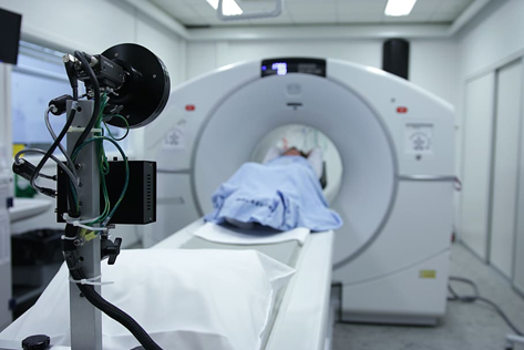
In order to better understand the complexity of the human brain, neuroimaging techniques have revolutionized the area of brain study. Neuroimaging technology has significantly improved over time, which has improved our comprehension of the connectivity, structure, and function of the brain.
Our everyday behavior and cognitive abilities are supported by the vast, complex, and dynamic human brain. For many years, scientists have been studying the mechanics of the human brain in both healthy and diseased states. Advanced neuroimaging methods, including magnetic resonance imaging (MRI), event-related potentials (ERPs), magnetoencephalography (MEG), and functional near-infrared spectroscopy (fNIRS), have been widely used to investigate the fundamentals of brain pathology changes in different brain disorders as well as the structural and functional architectures of the brain.
In particular, compared to single-modal neuroimaging techniques, multi-modal neuroimaging techniques will eventually offer a more thorough understanding of the pathomechanism of brain illnesses. This information can be helpful for early diagnosis, prognostication, and therapy effect evaluation. Generally speaking, the creation of reliable analysis techniques for neuroimaging will significantly improve our understanding of the brain and enable a multitude of neuroscience and clinical research projects. The most recent advancements in neuroimaging techniques are examined, emphasizing their potential to shed light on the mysteries of the human mind.
1. Magnetic Resonance Imaging (MRI):

For many years, MRI has been a pillar of brain research. Faster scan times and better spatial resolution are the results of recent MRI technological developments. Researchers can now map brain activity more precisely thanks to high-resolution functional MRI (fMRI), which reveals the complex networks that underlie cognitive activities like attention, memory, and decision-making.
2. Diffusion Tensor Imaging (DTI):
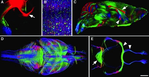
DTI is a specialized type of MRI that assesses the water molecule diffusion within the brain tissues. With knowledge about the white matter tracts in the brain, this method analyzes the connection and integrity of neuronal networks. Recent advancements in DTI recreate intricate fiber connections, helping researchers better understand neurodegenerative illnesses, traumatic brain injuries, and neurodevelopmental problems.
3. Positron Emission Tomography (PET):
In order to see brain activity, PET imaging entails injecting a radioactive tracer into the blood. Novel tracers that target certain neurotransmitter systems, receptors, and disease indicators are the results of advances in PET technology. It has become easier to study a variety of mental diseases, including Alzheimer’s disease, depression, and schizophrenia with neurotransmitter imbalances and disease progression information.
4. Electroencephalography (EEG):
By capturing electrical signals from the scalp, EEG analyses the electrical activity of the brain. Higher channel counts and more mobility are the results of recent advancements in EEG technology. Research into brain dynamics, neural oscillations, and brain-computer interfaces has been made possible by the improved extraction of relevant information from EEG data made possible by modern signal processing techniques. The potential of EEG in the diagnosis and monitoring of neurological illnesses, such as epilepsy and sleep disorders, is also being investigated.

5. Functional Near-Infrared Spectroscopy (fNIRS):
A non-invasive neuroimaging method called fNIRS monitors changes in the brain’s blood oxygenation levels. The introduction of wearable, wireless devices and better spatial resolution are both results of recent developments in fNIRS technology. For investigating infants, kids, and patients with movement constraints, fNIRS is particularly well suited. It is a promising substitute for existing imaging modalities and has applications in cognitive neuroscience, clinical research, and brain-computer interfaces.
6. Magnetoencephalography (MEG):
The magnetic fields produced by neural activity are measured by MEG. MEG technology has recently made strides that have boosted sensor density and reduced noise levels. This makes it possible to record the temporal dynamics of brain activity with accuracy, improving their comprehension of neural processes. MEG has shown to be helpful in understanding brain conditions. Conditions may include epilepsy and autism and has the potential to advance precision medicine by directing individualized therapies.
7. Optical Coherence Tomography (OCT):
OCT is a new method of neuroimaging that uses light waves to take detailed pictures of the brain. Optical Coherence Tomography imaging depth and speed have recently improved. It allows researchers to examine the microstructure of the brain, including neural networks and blood flow. Advanced neuroimaging technique shed light on the fundamental mechanisms causing neurovascular coupling, neurodegenerative disorders, and brain tumors.
Non-invasive Neuroimaging Techniques – Overview
Neuroimaging, often known as brain imaging techniques, is a non-invasive approach to examining brain structure and activity. Without requiring invasive neurosurgery, these methods enable scientists and medical professionals to view the brain and identify anomalies or modifications in brain activity.
One of the methods for brain imaging that is most frequently utilized is functional magnetic resonance imaging (fMRI). By observing variations in blood flow and oxygen delivery in response to neural activity, it gauges brain activity. This method can identify particular brain regions that are active during particular tasks or experiences and has a high spatial resolution. It is employed in the research of mental and neurological disorders, including as schizophrenia, anxiety, and depression.
Another method for creating 2D and 3D images of the brain using X-rays is called computed tomography (CT) scanning. Important brain characteristics including tumours, haemorrhages, and other structural anomalies can be seen on CT scans. Nevertheless, CT scans are not commonly utilised to research brain activity and are less useful in clarifying the structure of the brain.
Short-lived radiotracers are used in Positron Emission Tomography (PET), a brain imaging technique, to map the functional processes of the brain. Brain activity is correlated with areas of elevated radioactivity. PET is frequently used to diagnose Parkinson’s disease, Alzheimer’s disease, and other neurological illnesses. It is also used to investigate brain function and metabolism.
By recording from electrodes positioned on the scalp, electroencephalography (EEG) quantifies the electrical activity of the brain. An electrical signal from several neurons is represented by the electroencephalogram (EEG). EEG is widely utilised in research on sleep, memory, and other cognitive processes as well as in the diagnosis and monitoring of seizure disorders.
A kind of brain imaging called magnetoencephalography (MEG) gauges the magnetic fields produced by the brain’s electrical activity. This method detects the magnetic fields by means of sensitive instruments called SQUIDs (Superconducting Quantum Interference Devices). MEG is utilized to examine brain function in both healthy persons and patients with neurological diseases because of its great temporal resolution.
An optical brain imaging method called functional near-infrared spectroscopy (fNIRS) tracks variations in the blood oxygenation of the brain. fNIRS can be used to research brain activity in newborns and early children because of its great temporal resolution. Another optical method for studying retinal disorders is optical coherence tomography (OCT), which produces high-resolution brain images.
Learn Neuroimaging Techniques
Neuroanatomy, relevant experimental design concerns, knowledge of MRI scanning sequences, and a significant amount of computational competence are all necessary for successful use of neuroimaging. Particularly, when it comes to choosing and using the right processing software and interpreting the results. Currently, researchers interested in using and learning neuroimaging techniques in their field must make a significant effort to learn the necessary skills outside of their field of study. Efforts may not be worthwhile if preceed without receiving formal instruction. Various certificates program offer direct routes for gaining neuroimaging expertise, which frequently lies outside of the student’s primary field of study. You can learn neuroimaging techniques easily, clearly and professionally at Udemy and Coursera. Check out this link, perhaps you’d join these classes and step in to get an amazing exposure of neuroimaging techniques learning.
Check out and learn the neuroimaging principles and practice analysis skills in small classes with individual attention.
Wrapping Up:
Insights into the intricate workings of the human brain have never been more accessible thanks to developments in neuroimaging methods. Each technique adds a different perspective and completes other modalities in the study of brain anatomy, function, and connection, from MRI and DTI through PET, EEG, firs, MEG, and OCT. These developments have the potential to make significant progress in neuroscience. Also, such developments help us understand the mysteries of the mind, and ultimately diagnose and treat brain-related ailments better. We may anticipate more advances in neuroimaging methods as technology advances. These methods will offer up new vistas for research into the amazing organ that is the human brain.


